Parathyroid Glands provides an educational overview of calcium regulation, parathyroid hormone, anatomy, testing, and common disorders. It is designed for anatomy and terminology learning and cannot diagnose symptoms or replace professional medical care.
Essential definition
Parathyroid glands is best understood through the relationship between structure and secretion. A gland is an organized collection of cells that produces one or more substances and releases them either into the bloodstream or through a duct. Those substances may include hormones, enzymes, mucus, sweat, tears, saliva, oil, milk, or digestive fluid. The adjective adenous is a rare way of saying glandular or gland-like, but modern medical writing more often uses glandular, adenomatous, or a specific anatomical name.
Medical discussions become clearer when endocrine and exocrine functions are separated. Endocrine tissue releases hormones into the blood, allowing chemical signals to reach distant organs. Exocrine tissue usually releases its product through a duct onto an epithelial surface or into a body cavity. Some organs, especially the pancreas, contain both endocrine and exocrine components, so a single organ can participate in two very different forms of secretion.
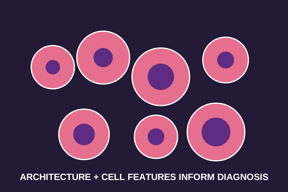
The microscopic architecture of glandular tissue often includes secretory units, ducts, blood vessels, supporting connective tissue, nerves, and immune cells. Depending on the organ, secretory cells may form acini, follicles, tubules, cords, nests, or lobules. A pathologist evaluates both individual cell features and the larger architectural pattern because normal function and disease can alter either level.
Secretion is an active biological process. Cells synthesize products, package or transport them, respond to signals, and release them in a controlled manner. Protein and peptide hormones may be stored in vesicles before release, while steroid hormones are synthesized from cholesterol and diffuse across cell membranes. Exocrine products may be released by exocytosis, partial loss of the cell apex, or complete breakdown of the secretory cell, depending on the gland.
Anatomy and organization
Feedback regulation helps keep many glandular systems within a functional range. A hormone released by one gland may stimulate another gland, and the final hormone may then inhibit the earlier steps. This negative-feedback arrangement is common in endocrine physiology. Timing matters: some hormones vary by hour, sleep, meals, stress, age, menstrual cycle, medication exposure, and illness, so one laboratory value is rarely interpreted without context.
Symptoms associated with gland disorders are often nonspecific. Fatigue, weight change, pain, swelling, skin changes, altered thirst, temperature intolerance, digestive symptoms, or reproductive changes can arise from many unrelated causes. A symptom does not identify a gland or diagnosis by itself. Clinicians combine history, examination, laboratory data, imaging, and sometimes tissue sampling before reaching a conclusion.
Laboratory testing should be interpreted using the correct reference interval, specimen type, timing, and clinical question. A result marked high or low is not automatically a diagnosis. Reference ranges differ among laboratories, and some tests are intended for screening while others are used for confirmation or monitoring. Hormone systems are particularly dependent on feedback loops, so paired measurements may be more informative than one isolated value.
Imaging can describe a gland's size, location, internal structure, blood flow, ducts, surrounding tissues, and focal lesions. Ultrasound is commonly used for superficial glands and fluid-filled structures. CT can provide rapid cross-sectional anatomy, MRI offers excellent soft-tissue contrast, and nuclear medicine can demonstrate function or uptake. The best study depends on the organ and the question being asked.
Cells and microscopic structure
Terms such as nodule, cyst, enlargement, hyperplasia, adenoma, dysplasia, and carcinoma are not interchangeable. A nodule is a descriptive finding. A cyst contains fluid or semisolid material. Hyperplasia means increased cell number. An adenoma is generally a benign glandular neoplasm. Dysplasia describes abnormal cellular development, and carcinoma is malignant epithelial disease. Location and pathology determine the actual significance.
Benign does not always mean unimportant. A noncancerous glandular lesion can produce excess hormone, compress nearby structures, obstruct a duct, bleed, become infected, or create cosmetic and functional problems. Conversely, many incidental nodules remain stable and never require treatment. Risk assessment depends on size, growth, symptoms, hormone activity, imaging features, age, family history, and pathology.
Inflammation may result from infection, autoimmune disease, obstruction, trauma, medication, radiation, or an unknown trigger. Medical terms often add the suffix -itis to the gland name, such as thyroiditis, pancreatitis, or sialadenitis. Inflammation can be acute or chronic and may temporarily increase, decrease, or otherwise alter secretion.
Tumors arising in glandular tissue are classified by organ, cell type, architecture, molecular features, grade, and stage. The prefix adeno- signals glandular origin or gland-like formation, but it does not by itself reveal whether a lesion is benign or malignant. Adenoma typically refers to a benign neoplasm, whereas adenocarcinoma refers to a malignant epithelial tumor showing glandular differentiation.
| Term | General meaning |
|---|---|
| Endocrine | Secretion into blood |
| Exocrine | Secretion through a duct or opening |
| Hyperplasia | Increase in cell number |
| Adenoma | Usually benign glandular neoplasm |
| Adenocarcinoma | Malignant tumor with glandular differentiation |
How secretion works
Treatment follows the cause rather than the word glandular. Options may include observation, symptom management, hormone replacement, medication that suppresses secretion, antibiotics when bacterial infection is present, drainage of an abscess or obstructed duct, image-guided procedures, surgery, radiation, systemic therapy, or combinations of these approaches. Decisions should be individualized by qualified clinicians.
A useful medical appointment begins with a focused record. Patients can note symptom timing, medication and supplement use, prior imaging, family history, recent infections, pregnancy status when relevant, and changes in weight, thirst, urination, temperature tolerance, skin, digestion, or menstrual function. Bringing prior reports rather than relying on memory can prevent duplicated testing and improve comparison over time.
Educational pages cannot determine whether a personal symptom is harmless or urgent. Rapid swelling, severe pain, trouble breathing or swallowing, fainting, confusion, severe dehydration, sudden weakness, uncontrolled bleeding, or symptoms of a major hormone crisis require prompt medical evaluation. The appropriate response depends on the complete clinical picture, not merely on the anatomical term involved.
The central lesson of parathyroid glands is that glandular anatomy, secretion, and disease must be described precisely. Readers should distinguish endocrine from exocrine tissue, structure from function, descriptive imaging terms from diagnoses, and benign growth from malignant disease. Reliable interpretation connects symptoms, testing, imaging, pathology, and clinical context.
Control and feedback
Parathyroid glands is best understood through the relationship between structure and secretion. A gland is an organized collection of cells that produces one or more substances and releases them either into the bloodstream or through a duct. Those substances may include hormones, enzymes, mucus, sweat, tears, saliva, oil, milk, or digestive fluid. The adjective adenous is a rare way of saying glandular or gland-like, but modern medical writing more often uses glandular, adenomatous, or a specific anatomical name.
Medical discussions become clearer when endocrine and exocrine functions are separated. Endocrine tissue releases hormones into the blood, allowing chemical signals to reach distant organs. Exocrine tissue usually releases its product through a duct onto an epithelial surface or into a body cavity. Some organs, especially the pancreas, contain both endocrine and exocrine components, so a single organ can participate in two very different forms of secretion.
The microscopic architecture of glandular tissue often includes secretory units, ducts, blood vessels, supporting connective tissue, nerves, and immune cells. Depending on the organ, secretory cells may form acini, follicles, tubules, cords, nests, or lobules. A pathologist evaluates both individual cell features and the larger architectural pattern because normal function and disease can alter either level.
Secretion is an active biological process. Cells synthesize products, package or transport them, respond to signals, and release them in a controlled manner. Protein and peptide hormones may be stored in vesicles before release, while steroid hormones are synthesized from cholesterol and diffuse across cell membranes. Exocrine products may be released by exocytosis, partial loss of the cell apex, or complete breakdown of the secretory cell, depending on the gland.
Development and variation
Feedback regulation helps keep many glandular systems within a functional range. A hormone released by one gland may stimulate another gland, and the final hormone may then inhibit the earlier steps. This negative-feedback arrangement is common in endocrine physiology. Timing matters: some hormones vary by hour, sleep, meals, stress, age, menstrual cycle, medication exposure, and illness, so one laboratory value is rarely interpreted without context.
Symptoms associated with gland disorders are often nonspecific. Fatigue, weight change, pain, swelling, skin changes, altered thirst, temperature intolerance, digestive symptoms, or reproductive changes can arise from many unrelated causes. A symptom does not identify a gland or diagnosis by itself. Clinicians combine history, examination, laboratory data, imaging, and sometimes tissue sampling before reaching a conclusion.
Laboratory testing should be interpreted using the correct reference interval, specimen type, timing, and clinical question. A result marked high or low is not automatically a diagnosis. Reference ranges differ among laboratories, and some tests are intended for screening while others are used for confirmation or monitoring. Hormone systems are particularly dependent on feedback loops, so paired measurements may be more informative than one isolated value.
Imaging can describe a gland's size, location, internal structure, blood flow, ducts, surrounding tissues, and focal lesions. Ultrasound is commonly used for superficial glands and fluid-filled structures. CT can provide rapid cross-sectional anatomy, MRI offers excellent soft-tissue contrast, and nuclear medicine can demonstrate function or uptake. The best study depends on the organ and the question being asked.
Common symptoms and clinical questions
Terms such as nodule, cyst, enlargement, hyperplasia, adenoma, dysplasia, and carcinoma are not interchangeable. A nodule is a descriptive finding. A cyst contains fluid or semisolid material. Hyperplasia means increased cell number. An adenoma is generally a benign glandular neoplasm. Dysplasia describes abnormal cellular development, and carcinoma is malignant epithelial disease. Location and pathology determine the actual significance.
Benign does not always mean unimportant. A noncancerous glandular lesion can produce excess hormone, compress nearby structures, obstruct a duct, bleed, become infected, or create cosmetic and functional problems. Conversely, many incidental nodules remain stable and never require treatment. Risk assessment depends on size, growth, symptoms, hormone activity, imaging features, age, family history, and pathology.
Inflammation may result from infection, autoimmune disease, obstruction, trauma, medication, radiation, or an unknown trigger. Medical terms often add the suffix -itis to the gland name, such as thyroiditis, pancreatitis, or sialadenitis. Inflammation can be acute or chronic and may temporarily increase, decrease, or otherwise alter secretion.
Tumors arising in glandular tissue are classified by organ, cell type, architecture, molecular features, grade, and stage. The prefix adeno- signals glandular origin or gland-like formation, but it does not by itself reveal whether a lesion is benign or malignant. Adenoma typically refers to a benign neoplasm, whereas adenocarcinoma refers to a malignant epithelial tumor showing glandular differentiation.
| Term | General meaning |
|---|---|
| Endocrine | Secretion into blood |
| Exocrine | Secretion through a duct or opening |
| Hyperplasia | Increase in cell number |
| Adenoma | Usually benign glandular neoplasm |
| Adenocarcinoma | Malignant tumor with glandular differentiation |
Testing and evaluation
Treatment follows the cause rather than the word glandular. Options may include observation, symptom management, hormone replacement, medication that suppresses secretion, antibiotics when bacterial infection is present, drainage of an abscess or obstructed duct, image-guided procedures, surgery, radiation, systemic therapy, or combinations of these approaches. Decisions should be individualized by qualified clinicians.
A useful medical appointment begins with a focused record. Patients can note symptom timing, medication and supplement use, prior imaging, family history, recent infections, pregnancy status when relevant, and changes in weight, thirst, urination, temperature tolerance, skin, digestion, or menstrual function. Bringing prior reports rather than relying on memory can prevent duplicated testing and improve comparison over time.
Educational pages cannot determine whether a personal symptom is harmless or urgent. Rapid swelling, severe pain, trouble breathing or swallowing, fainting, confusion, severe dehydration, sudden weakness, uncontrolled bleeding, or symptoms of a major hormone crisis require prompt medical evaluation. The appropriate response depends on the complete clinical picture, not merely on the anatomical term involved.
The central lesson of parathyroid glands is that glandular anatomy, secretion, and disease must be described precisely. Readers should distinguish endocrine from exocrine tissue, structure from function, descriptive imaging terms from diagnoses, and benign growth from malignant disease. Reliable interpretation connects symptoms, testing, imaging, pathology, and clinical context.
Imaging and procedures
Parathyroid glands is best understood through the relationship between structure and secretion. A gland is an organized collection of cells that produces one or more substances and releases them either into the bloodstream or through a duct. Those substances may include hormones, enzymes, mucus, sweat, tears, saliva, oil, milk, or digestive fluid. The adjective adenous is a rare way of saying glandular or gland-like, but modern medical writing more often uses glandular, adenomatous, or a specific anatomical name.
Medical discussions become clearer when endocrine and exocrine functions are separated. Endocrine tissue releases hormones into the blood, allowing chemical signals to reach distant organs. Exocrine tissue usually releases its product through a duct onto an epithelial surface or into a body cavity. Some organs, especially the pancreas, contain both endocrine and exocrine components, so a single organ can participate in two very different forms of secretion.
The microscopic architecture of glandular tissue often includes secretory units, ducts, blood vessels, supporting connective tissue, nerves, and immune cells. Depending on the organ, secretory cells may form acini, follicles, tubules, cords, nests, or lobules. A pathologist evaluates both individual cell features and the larger architectural pattern because normal function and disease can alter either level.
Secretion is an active biological process. Cells synthesize products, package or transport them, respond to signals, and release them in a controlled manner. Protein and peptide hormones may be stored in vesicles before release, while steroid hormones are synthesized from cholesterol and diffuse across cell membranes. Exocrine products may be released by exocytosis, partial loss of the cell apex, or complete breakdown of the secretory cell, depending on the gland.
Benign conditions
Feedback regulation helps keep many glandular systems within a functional range. A hormone released by one gland may stimulate another gland, and the final hormone may then inhibit the earlier steps. This negative-feedback arrangement is common in endocrine physiology. Timing matters: some hormones vary by hour, sleep, meals, stress, age, menstrual cycle, medication exposure, and illness, so one laboratory value is rarely interpreted without context.
Symptoms associated with gland disorders are often nonspecific. Fatigue, weight change, pain, swelling, skin changes, altered thirst, temperature intolerance, digestive symptoms, or reproductive changes can arise from many unrelated causes. A symptom does not identify a gland or diagnosis by itself. Clinicians combine history, examination, laboratory data, imaging, and sometimes tissue sampling before reaching a conclusion.
Laboratory testing should be interpreted using the correct reference interval, specimen type, timing, and clinical question. A result marked high or low is not automatically a diagnosis. Reference ranges differ among laboratories, and some tests are intended for screening while others are used for confirmation or monitoring. Hormone systems are particularly dependent on feedback loops, so paired measurements may be more informative than one isolated value.
Imaging can describe a gland's size, location, internal structure, blood flow, ducts, surrounding tissues, and focal lesions. Ultrasound is commonly used for superficial glands and fluid-filled structures. CT can provide rapid cross-sectional anatomy, MRI offers excellent soft-tissue contrast, and nuclear medicine can demonstrate function or uptake. The best study depends on the organ and the question being asked.
Inflammation and infection
Terms such as nodule, cyst, enlargement, hyperplasia, adenoma, dysplasia, and carcinoma are not interchangeable. A nodule is a descriptive finding. A cyst contains fluid or semisolid material. Hyperplasia means increased cell number. An adenoma is generally a benign glandular neoplasm. Dysplasia describes abnormal cellular development, and carcinoma is malignant epithelial disease. Location and pathology determine the actual significance.
Benign does not always mean unimportant. A noncancerous glandular lesion can produce excess hormone, compress nearby structures, obstruct a duct, bleed, become infected, or create cosmetic and functional problems. Conversely, many incidental nodules remain stable and never require treatment. Risk assessment depends on size, growth, symptoms, hormone activity, imaging features, age, family history, and pathology.
Inflammation may result from infection, autoimmune disease, obstruction, trauma, medication, radiation, or an unknown trigger. Medical terms often add the suffix -itis to the gland name, such as thyroiditis, pancreatitis, or sialadenitis. Inflammation can be acute or chronic and may temporarily increase, decrease, or otherwise alter secretion.
Tumors arising in glandular tissue are classified by organ, cell type, architecture, molecular features, grade, and stage. The prefix adeno- signals glandular origin or gland-like formation, but it does not by itself reveal whether a lesion is benign or malignant. Adenoma typically refers to a benign neoplasm, whereas adenocarcinoma refers to a malignant epithelial tumor showing glandular differentiation.
| Term | General meaning |
|---|---|
| Endocrine | Secretion into blood |
| Exocrine | Secretion through a duct or opening |
| Hyperplasia | Increase in cell number |
| Adenoma | Usually benign glandular neoplasm |
| Adenocarcinoma | Malignant tumor with glandular differentiation |
Tumors and abnormal growth
Treatment follows the cause rather than the word glandular. Options may include observation, symptom management, hormone replacement, medication that suppresses secretion, antibiotics when bacterial infection is present, drainage of an abscess or obstructed duct, image-guided procedures, surgery, radiation, systemic therapy, or combinations of these approaches. Decisions should be individualized by qualified clinicians.
A useful medical appointment begins with a focused record. Patients can note symptom timing, medication and supplement use, prior imaging, family history, recent infections, pregnancy status when relevant, and changes in weight, thirst, urination, temperature tolerance, skin, digestion, or menstrual function. Bringing prior reports rather than relying on memory can prevent duplicated testing and improve comparison over time.
Educational pages cannot determine whether a personal symptom is harmless or urgent. Rapid swelling, severe pain, trouble breathing or swallowing, fainting, confusion, severe dehydration, sudden weakness, uncontrolled bleeding, or symptoms of a major hormone crisis require prompt medical evaluation. The appropriate response depends on the complete clinical picture, not merely on the anatomical term involved.
The central lesson of parathyroid glands is that glandular anatomy, secretion, and disease must be described precisely. Readers should distinguish endocrine from exocrine tissue, structure from function, descriptive imaging terms from diagnoses, and benign growth from malignant disease. Reliable interpretation connects symptoms, testing, imaging, pathology, and clinical context.
Treatment principles
Parathyroid glands is best understood through the relationship between structure and secretion. A gland is an organized collection of cells that produces one or more substances and releases them either into the bloodstream or through a duct. Those substances may include hormones, enzymes, mucus, sweat, tears, saliva, oil, milk, or digestive fluid. The adjective adenous is a rare way of saying glandular or gland-like, but modern medical writing more often uses glandular, adenomatous, or a specific anatomical name.
Medical discussions become clearer when endocrine and exocrine functions are separated. Endocrine tissue releases hormones into the blood, allowing chemical signals to reach distant organs. Exocrine tissue usually releases its product through a duct onto an epithelial surface or into a body cavity. Some organs, especially the pancreas, contain both endocrine and exocrine components, so a single organ can participate in two very different forms of secretion.
The microscopic architecture of glandular tissue often includes secretory units, ducts, blood vessels, supporting connective tissue, nerves, and immune cells. Depending on the organ, secretory cells may form acini, follicles, tubules, cords, nests, or lobules. A pathologist evaluates both individual cell features and the larger architectural pattern because normal function and disease can alter either level.
Secretion is an active biological process. Cells synthesize products, package or transport them, respond to signals, and release them in a controlled manner. Protein and peptide hormones may be stored in vesicles before release, while steroid hormones are synthesized from cholesterol and diffuse across cell membranes. Exocrine products may be released by exocytosis, partial loss of the cell apex, or complete breakdown of the secretory cell, depending on the gland.
Questions to ask a clinician
Feedback regulation helps keep many glandular systems within a functional range. A hormone released by one gland may stimulate another gland, and the final hormone may then inhibit the earlier steps. This negative-feedback arrangement is common in endocrine physiology. Timing matters: some hormones vary by hour, sleep, meals, stress, age, menstrual cycle, medication exposure, and illness, so one laboratory value is rarely interpreted without context.
Symptoms associated with gland disorders are often nonspecific. Fatigue, weight change, pain, swelling, skin changes, altered thirst, temperature intolerance, digestive symptoms, or reproductive changes can arise from many unrelated causes. A symptom does not identify a gland or diagnosis by itself. Clinicians combine history, examination, laboratory data, imaging, and sometimes tissue sampling before reaching a conclusion.
Laboratory testing should be interpreted using the correct reference interval, specimen type, timing, and clinical question. A result marked high or low is not automatically a diagnosis. Reference ranges differ among laboratories, and some tests are intended for screening while others are used for confirmation or monitoring. Hormone systems are particularly dependent on feedback loops, so paired measurements may be more informative than one isolated value.
Imaging can describe a gland's size, location, internal structure, blood flow, ducts, surrounding tissues, and focal lesions. Ultrasound is commonly used for superficial glands and fluid-filled structures. CT can provide rapid cross-sectional anatomy, MRI offers excellent soft-tissue contrast, and nuclear medicine can demonstrate function or uptake. The best study depends on the organ and the question being asked.
Common misunderstandings
Terms such as nodule, cyst, enlargement, hyperplasia, adenoma, dysplasia, and carcinoma are not interchangeable. A nodule is a descriptive finding. A cyst contains fluid or semisolid material. Hyperplasia means increased cell number. An adenoma is generally a benign glandular neoplasm. Dysplasia describes abnormal cellular development, and carcinoma is malignant epithelial disease. Location and pathology determine the actual significance.
Benign does not always mean unimportant. A noncancerous glandular lesion can produce excess hormone, compress nearby structures, obstruct a duct, bleed, become infected, or create cosmetic and functional problems. Conversely, many incidental nodules remain stable and never require treatment. Risk assessment depends on size, growth, symptoms, hormone activity, imaging features, age, family history, and pathology.
Inflammation may result from infection, autoimmune disease, obstruction, trauma, medication, radiation, or an unknown trigger. Medical terms often add the suffix -itis to the gland name, such as thyroiditis, pancreatitis, or sialadenitis. Inflammation can be acute or chronic and may temporarily increase, decrease, or otherwise alter secretion.
Tumors arising in glandular tissue are classified by organ, cell type, architecture, molecular features, grade, and stage. The prefix adeno- signals glandular origin or gland-like formation, but it does not by itself reveal whether a lesion is benign or malignant. Adenoma typically refers to a benign neoplasm, whereas adenocarcinoma refers to a malignant epithelial tumor showing glandular differentiation.
| Term | General meaning |
|---|---|
| Endocrine | Secretion into blood |
| Exocrine | Secretion through a duct or opening |
| Hyperplasia | Increase in cell number |
| Adenoma | Usually benign glandular neoplasm |
| Adenocarcinoma | Malignant tumor with glandular differentiation |
Summary
Treatment follows the cause rather than the word glandular. Options may include observation, symptom management, hormone replacement, medication that suppresses secretion, antibiotics when bacterial infection is present, drainage of an abscess or obstructed duct, image-guided procedures, surgery, radiation, systemic therapy, or combinations of these approaches. Decisions should be individualized by qualified clinicians.
A useful medical appointment begins with a focused record. Patients can note symptom timing, medication and supplement use, prior imaging, family history, recent infections, pregnancy status when relevant, and changes in weight, thirst, urination, temperature tolerance, skin, digestion, or menstrual function. Bringing prior reports rather than relying on memory can prevent duplicated testing and improve comparison over time.
Educational pages cannot determine whether a personal symptom is harmless or urgent. Rapid swelling, severe pain, trouble breathing or swallowing, fainting, confusion, severe dehydration, sudden weakness, uncontrolled bleeding, or symptoms of a major hormone crisis require prompt medical evaluation. The appropriate response depends on the complete clinical picture, not merely on the anatomical term involved.
The central lesson of parathyroid glands is that glandular anatomy, secretion, and disease must be described precisely. Readers should distinguish endocrine from exocrine tissue, structure from function, descriptive imaging terms from diagnoses, and benign growth from malignant disease. Reliable interpretation connects symptoms, testing, imaging, pathology, and clinical context.
Essential definition
Parathyroid glands is best understood through the relationship between structure and secretion. A gland is an organized collection of cells that produces one or more substances and releases them either into the bloodstream or through a duct. Those substances may include hormones, enzymes, mucus, sweat, tears, saliva, oil, milk, or digestive fluid. The adjective adenous is a rare way of saying glandular or gland-like, but modern medical writing more often uses glandular, adenomatous, or a specific anatomical name.
Medical discussions become clearer when endocrine and exocrine functions are separated. Endocrine tissue releases hormones into the blood, allowing chemical signals to reach distant organs. Exocrine tissue usually releases its product through a duct onto an epithelial surface or into a body cavity. Some organs, especially the pancreas, contain both endocrine and exocrine components, so a single organ can participate in two very different forms of secretion.
The microscopic architecture of glandular tissue often includes secretory units, ducts, blood vessels, supporting connective tissue, nerves, and immune cells. Depending on the organ, secretory cells may form acini, follicles, tubules, cords, nests, or lobules. A pathologist evaluates both individual cell features and the larger architectural pattern because normal function and disease can alter either level.
Secretion is an active biological process. Cells synthesize products, package or transport them, respond to signals, and release them in a controlled manner. Protein and peptide hormones may be stored in vesicles before release, while steroid hormones are synthesized from cholesterol and diffuse across cell membranes. Exocrine products may be released by exocytosis, partial loss of the cell apex, or complete breakdown of the secretory cell, depending on the gland.
Anatomy and organization
Feedback regulation helps keep many glandular systems within a functional range. A hormone released by one gland may stimulate another gland, and the final hormone may then inhibit the earlier steps. This negative-feedback arrangement is common in endocrine physiology. Timing matters: some hormones vary by hour, sleep, meals, stress, age, menstrual cycle, medication exposure, and illness, so one laboratory value is rarely interpreted without context.
Symptoms associated with gland disorders are often nonspecific. Fatigue, weight change, pain, swelling, skin changes, altered thirst, temperature intolerance, digestive symptoms, or reproductive changes can arise from many unrelated causes. A symptom does not identify a gland or diagnosis by itself. Clinicians combine history, examination, laboratory data, imaging, and sometimes tissue sampling before reaching a conclusion.
Laboratory testing should be interpreted using the correct reference interval, specimen type, timing, and clinical question. A result marked high or low is not automatically a diagnosis. Reference ranges differ among laboratories, and some tests are intended for screening while others are used for confirmation or monitoring. Hormone systems are particularly dependent on feedback loops, so paired measurements may be more informative than one isolated value.
Imaging can describe a gland's size, location, internal structure, blood flow, ducts, surrounding tissues, and focal lesions. Ultrasound is commonly used for superficial glands and fluid-filled structures. CT can provide rapid cross-sectional anatomy, MRI offers excellent soft-tissue contrast, and nuclear medicine can demonstrate function or uptake. The best study depends on the organ and the question being asked.
Cells and microscopic structure
Terms such as nodule, cyst, enlargement, hyperplasia, adenoma, dysplasia, and carcinoma are not interchangeable. A nodule is a descriptive finding. A cyst contains fluid or semisolid material. Hyperplasia means increased cell number. An adenoma is generally a benign glandular neoplasm. Dysplasia describes abnormal cellular development, and carcinoma is malignant epithelial disease. Location and pathology determine the actual significance.
Benign does not always mean unimportant. A noncancerous glandular lesion can produce excess hormone, compress nearby structures, obstruct a duct, bleed, become infected, or create cosmetic and functional problems. Conversely, many incidental nodules remain stable and never require treatment. Risk assessment depends on size, growth, symptoms, hormone activity, imaging features, age, family history, and pathology.
Inflammation may result from infection, autoimmune disease, obstruction, trauma, medication, radiation, or an unknown trigger. Medical terms often add the suffix -itis to the gland name, such as thyroiditis, pancreatitis, or sialadenitis. Inflammation can be acute or chronic and may temporarily increase, decrease, or otherwise alter secretion.
Tumors arising in glandular tissue are classified by organ, cell type, architecture, molecular features, grade, and stage. The prefix adeno- signals glandular origin or gland-like formation, but it does not by itself reveal whether a lesion is benign or malignant. Adenoma typically refers to a benign neoplasm, whereas adenocarcinoma refers to a malignant epithelial tumor showing glandular differentiation.
| Term | General meaning |
|---|---|
| Endocrine | Secretion into blood |
| Exocrine | Secretion through a duct or opening |
| Hyperplasia | Increase in cell number |
| Adenoma | Usually benign glandular neoplasm |
| Adenocarcinoma | Malignant tumor with glandular differentiation |
How secretion works
Treatment follows the cause rather than the word glandular. Options may include observation, symptom management, hormone replacement, medication that suppresses secretion, antibiotics when bacterial infection is present, drainage of an abscess or obstructed duct, image-guided procedures, surgery, radiation, systemic therapy, or combinations of these approaches. Decisions should be individualized by qualified clinicians.
A useful medical appointment begins with a focused record. Patients can note symptom timing, medication and supplement use, prior imaging, family history, recent infections, pregnancy status when relevant, and changes in weight, thirst, urination, temperature tolerance, skin, digestion, or menstrual function. Bringing prior reports rather than relying on memory can prevent duplicated testing and improve comparison over time.
Educational pages cannot determine whether a personal symptom is harmless or urgent. Rapid swelling, severe pain, trouble breathing or swallowing, fainting, confusion, severe dehydration, sudden weakness, uncontrolled bleeding, or symptoms of a major hormone crisis require prompt medical evaluation. The appropriate response depends on the complete clinical picture, not merely on the anatomical term involved.
The central lesson of parathyroid glands is that glandular anatomy, secretion, and disease must be described precisely. Readers should distinguish endocrine from exocrine tissue, structure from function, descriptive imaging terms from diagnoses, and benign growth from malignant disease. Reliable interpretation connects symptoms, testing, imaging, pathology, and clinical context.
Control and feedback
Parathyroid glands is best understood through the relationship between structure and secretion. A gland is an organized collection of cells that produces one or more substances and releases them either into the bloodstream or through a duct. Those substances may include hormones, enzymes, mucus, sweat, tears, saliva, oil, milk, or digestive fluid. The adjective adenous is a rare way of saying glandular or gland-like, but modern medical writing more often uses glandular, adenomatous, or a specific anatomical name.
Medical discussions become clearer when endocrine and exocrine functions are separated. Endocrine tissue releases hormones into the blood, allowing chemical signals to reach distant organs. Exocrine tissue usually releases its product through a duct onto an epithelial surface or into a body cavity. Some organs, especially the pancreas, contain both endocrine and exocrine components, so a single organ can participate in two very different forms of secretion.
The microscopic architecture of glandular tissue often includes secretory units, ducts, blood vessels, supporting connective tissue, nerves, and immune cells. Depending on the organ, secretory cells may form acini, follicles, tubules, cords, nests, or lobules. A pathologist evaluates both individual cell features and the larger architectural pattern because normal function and disease can alter either level.
Secretion is an active biological process. Cells synthesize products, package or transport them, respond to signals, and release them in a controlled manner. Protein and peptide hormones may be stored in vesicles before release, while steroid hormones are synthesized from cholesterol and diffuse across cell membranes. Exocrine products may be released by exocytosis, partial loss of the cell apex, or complete breakdown of the secretory cell, depending on the gland.
Frequently asked questions
What does adenous mean?
Adenous is a rare adjective meaning glandular, gland-like, or relating to a gland.
Is adenous a common medical term?
No. Modern medical writing more commonly uses glandular or a specific anatomical or pathological term.
What is a gland?
A gland is an organ or organized group of cells that makes and releases one or more substances.
What is the difference between endocrine and exocrine glands?
Endocrine glands release hormones into the blood; exocrine glands release products through ducts or openings.
Is an adenoma cancer?
An adenoma is generally a benign glandular neoplasm, although some adenomas can require monitoring or treatment.
What is adenocarcinoma?
Adenocarcinoma is a malignant epithelial tumor that forms glands or arises from glandular cells.
Does a gland nodule mean cancer?
No. Nodule is a descriptive term, and many gland nodules are benign.
Why do gland tests vary by time of day?
Some hormones follow daily rhythms or change with meals, stress, sleep, medications, and other conditions.
Can a gland be both endocrine and exocrine?
Yes. The pancreas has endocrine islets and exocrine acinar and duct tissue.
Can online information diagnose a gland disorder?
No. Diagnosis requires clinical evaluation and sometimes laboratory, imaging, or pathology findings.